
Dental X-Rays and Radiation: A Patient Guide
.avif)
Patients may wonder why imaging is necessary when the visible teeth appear healthy. This guide explains why dental X-rays are necessary and how dentists select imaging, manage exposure, and interpret results.
Why Dental X-Rays Matter in Dental Care
An oral examination cannot show every surface, tooth root, restoration margin, or area of supporting bone; FDA dental radiographic guidance notes that radiographs can help identify caries and periodontal bone loss that may not be apparent clinically. According to the American Dental Association, X-rays can help assess dental disease, bone health, tooth roots, eruption patterns, and treatment planning when prescribed according to clinical need. For children, dental imaging is especially important to monitor tooth eruption and development. A full-mouth series may be recommended when a comprehensive view of all teeth and supporting structures is needed, particularly in children's dental imaging to track growth and identify any abnormalities early.
Imaging frequency should reflect symptoms, examination findings, oral-health history, and individual risk factors rather than a universal schedule. The ADA/FDA patient-selection recommendations advise bitewing X-rays every 24–36 months for adults with no clinical issues and no increased risk, and every 6–18 months for adults with increased risk factors, while leaving the type, frequency, and extent of imaging to professional judgment.
What Dentists Can and Cannot See Without Imaging
A dentist can inspect exposed teeth and soft tissues, but surfaces in between the teeth and structures below the gums may remain hidden. Imaging contributes to an accurate diagnosis alongside clinical examination, patient history, and symptoms.
Understanding Radiation From Dental X-Rays
context for the everyday radiation exposure humans receivTo understand the level of radiation emitted by dental X-rays, it is important to have the context of everyday radiation exposure humans receive in daily life. The graphic below compares radiation exposure from dental X-rays with that from daily activities.
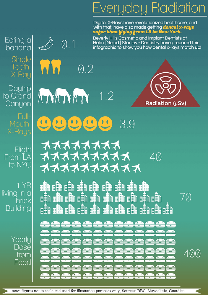
Why Exposure Varies
Radiation exposure varies with image type, equipment, settings, number of views, and patient size. The IAEA's dental-radiology dose guidance reports typical effective doses of about 1–8 µSv for intraoral X-rays, approximately 4–30 µSv for panoramic examinations, and roughly 50 µSv or below for small- or medium-field CBCT scans, with larger CBCT fields around 100 µSv or more.
How Digital Dental Imaging Supports Dose Reduction
Digital X-rays include intraoral X-rays, panoramic X-rays, and specialized systems with distinct purposes. FDA guidance on digital imaging states that digital imaging can reduce dose by 40–60% compared with film, depending on the receptor type and protocol; Dentists are instructed to use exposure that is as low as reasonably achievable while producing clinically useful information.
Choosing the Right Image for the Question
Bitewings commonly evaluate areas between teeth, while other images show an entire tooth and surrounding structures. A full-mouth series consists of multiple intraoral X-rays that together provide a detailed view of all teeth and the supporting bone, making it valuable for dental imaging in both adults and children. Panoramic images provide a broad overview, whereas cone-beam computed tomography, or CBCT, may be considered when three-dimensional detail could change diagnosis or treatment planning.
What to Expect During a Gainesville X-Ray Appointment
The dental team at Weber Dental reviews the relevant history, explains the recommended imaging, positions an intra-oral sensor or an external scanner, captures the views, and discusses significant findings. Before imaging begins, disclose pregnancy or possible pregnancy, previous imaging, medical concerns, and any questions about exposure.
Patients may request an explanation of benefits and alternatives, as well as confirmation of whether existing images remain diagnostically useful.
Questions to Ask Before Imaging
- Why is this X-ray needed today, and what will it evaluate?
- Are prior images available for review?
- Which imaging type is most appropriate for my circumstances?
- Could the result change diagnosis or treatment planning?
Common Misunderstandings About Dental X-Rays and Radiation
Dental examinations do not all involve the same radiation dose or produce the same diagnostic information. Declining clinically indicated imaging may leave inter-tooth decay, infection, bone changes, or root concerns undetected, although the benefits and limitations require individualized discussion rather than broad safety guarantees.
When to Seek Personalized Guidance
Pregnant patients, children, people with previous radiation exposure, and those with complex medical histories should discuss imaging with their treating dentist and, when appropriate, another qualified healthcare professional. This guide provides general education and does not replace individualized medical or dental advice.
Frequently Asked Questions
Is Radiation Exposure From Dental X-Rays Harmful?
X-rays use ionizing radiation, so each examination should be justified and optimized. A dentist can explain the expected diagnostic benefit, relevant limitations, and available alternatives for your situation.
How Much Radiation Will I Receive From Dental X-Rays?
We recommend X-rays once a year. It's usually four to eight X rays. That's about 1.8 µSv, which is negligible.
Are Dental X-Rays Safer Now?
Digital systems can reduce dose compared with film when used with appropriate receptors and protocols. Responsible practice still depends on ordering imaging only when clinically indicated and using appropriate exposure settings.
Which Dental X-Ray Gives the Least Radiation?
The answer depends on the equipment and technique, while each image type addresses a different diagnostic question. The appropriate choice is the examination that obtains necessary information with properly optimized exposure.
Key Takeaways for Gainesville Patients
Dental X-rays help dentists evaluate structures that a standard visual examination cannot fully reveal. Modern digital imaging and exposure optimization support responsible decision-making, so ask your Gainesville dental team to explain the purpose and expected value of every recommended image.
Experience Exceptional Dental Care
Join the Weber Dental family of satisfied patients and discover your best smile!
